


PRODUCTS

APEXVIEW APX100 All-in-One Microscope
The APX100 microscope supports a wide range of research imaging applications for slides, dishes, and well plates. Use the included imaging methods such as multichannel, stitching, time-lapse, and Z-stack acquisition in any combination to fit your research protocol.
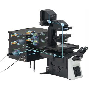
CONFOCAL LASER SCANNING MICROSCOPY
Laser scanning Confocal Microscopy (LSCM) is used in biological research to obtain high-resolution, high-contrast imagery of a sample. Laser microscopes can scan samples point by point, resulting in optical sectioning that can be used to construct precise 3D imagery. Laser scanning microscopes are designed with a large range of imaging modalities to meet some of the most difficult challenges in the life sciences. The FLUOVIEW FV3000 series of confocal laser scanning microscopes meets some of the most difficult challenges in modern science. Featuring the high sensitivity and speed required for live cell imaging as well as deep tissue observation, the FV3000 confocal microscope enables a wide range of imaging modalities, including macro-to-micro imaging, super resolution microscopy, and quantitative data analysis.
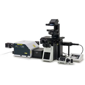
SPINNING DISK CONFOCAL MICROSCOPY (SDCM)
Spinning disk confocal microscopy (SDCM) represents an alternative to LSCM. The Spinning Disk unit helps you acquire high-speed confocal images with a larger field of view than traditional confocal microscopes. Deconvolution algorithms can be applied to improve image resolution, contrast, and dynamic range for strikingly clear 3D images, even at greater observation depths. The Yokogawa CSU-W1 spinning disk unit helps you acquire high-speed confocal images with a larger field of view than traditional confocal microscopes.
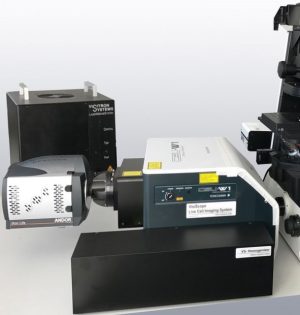

VisiScope Confocal
Real-Time Confocal for wide field of view, improved image quality and high speed live cell imaging

Stimulated emission depletion (STED) microscopy
The VisiScope-Real-Time Confocal System can be either based on Yokogawa CSU-X1 or CSU-W1 scan head.

stochastic optical reconstruction microscopy (STORM)
Stochastic Optical Reconstruction Microscopy (STORM) exploits the photoswitching properties of some fluorophores. In certain conditions, fluorophores can be sent to an intermediary state (the “dark state”) from which they randomly cycle between ON and OFF states.
In the proper chemical and excitation conditions the transition to the dark state is in the range of milliseconds to minutes and, importantly, is a completely stochastic event. As a result, when in the right chemical and excitation conditions, fluorophores start blinking randomly and in an asynchronous manner


cellTIRF
Total internal reflection fluorescence microscopy (TIRF) makes use of specific optics to produce illumination light only at the 50-100 nm range at the interface of the slide, massively reducing out of focus light and improving the ability to detect fluorescent molecules. Because of its low light intensity and high spatial resolution, it is a key technique in live-cell imaging. Designed for membrane dynamics, single molecule detection, and colocalization experiments, the IXplore TIRF microscope system offers simultaneous multicolor TIRF imaging for up to 4 colors with high stability. The Total Internal Reflection Fluorescence (TIRF) technique is the ideal method for observations of cells close to the coverslip surface.

Multiphoton Laser Scanning Microscope
Designed for deep imaging in biological specimens, the multiphoton microscope helps reveal how cells function and interact within living tissue.
Two-photon excitation microscopy (also called Multiphoton) is a fluorescence imaging technique that allows imaging of living tissue up to about one millimeter in thickness, Unlike traditional fluorescence microscopy, in which the excitation wavelength is shorter than the emission wavelength, two-photon excitation requires simultaneous excitation by two photons with longer wavelength than the emitted light. Two-photon excitation can be a superior alternative to confocal microscopy due to its deeper tissue penetration, efficient light detection, and reduced photobleaching.


Photocurrent Measuring Instruments
Photocurrent measurement is currently one of the hottest research topics in the material analysis field, especially in semiconductor and solar cell development industries.


27MG Ultrasonic Thickness Gauge
The 27MG basic ultrasonic thickness gauge is designed to make accurate measurements from one side on internally corroded or eroded metal pipes and parts. It is lightweight, durable, and ergonomically designed for easy, one-hand operation.
The Evident 27MG is an affordable ultrasonic thickness gauge designed to make accurate, measurements from one side on internally corroded or eroded metal pipes, tanks, and other equipment. It weighs only 12 oz. (340 g) and is ergonomically designed for easy, one-hand operation. Despite its compact size, the 27MG has many innovative measurement features utilizing the same technologies that are available on our more advanced thickness gauges. The durable, rugged 27MG is battery-operated and features a large, backlit LCD with easy-to-read numerals, and an intuitive color-coded keypad with direct access to many key features.

45MG Ultrasonic Thickness Gauge
The 45MG advanced ultrasonic thickness gauge is packed with standard measurement features and software options. This unique instrument is compatible with the complete range of Evident dual element and single element thickness gage transducers.
The 45MG is an advanced ultrasonic thickness gauge packed with standard measurement features and software options. This unique instrument is compatible with the complete range of Evident dual element and single element thickness gauge transducers, making this innovative instrument an all-in-one solution for virtually every thickness gauge application.