


PRODUCTS
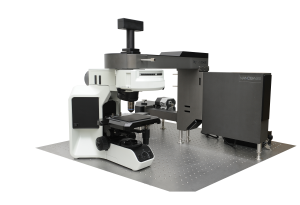
Raman Microscopy
NANOBASE provides the total solutions for Raman spectroscopy research. XPER MAKES YOUR RAMAN DREAMS COME TRUE

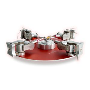
NanoWorkstation
Adds the ‘hand’ function to the electron microscope to allow physical manipulation and characterization at the micro- and nanoscale.
The module-based system offers high versatility, giving you the flexibility to perform numerous different specialized applications by simply changing the tool attached to the front of the manipulator, whether it is moving, assembling, preparing, rotating, pushing, probing, feeling, listening, gripping or any other task you can imagine.
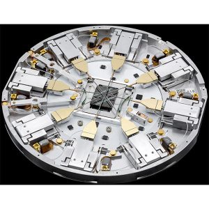

Probe Workstations
The most compact and highly integrated nanoprobing system in the market. With positional encoders on the platform, enables fast and efficient nanoprobing workflows.
Kleindiek Nanotechnik is dedicated to providing you with sophisticated and easy to use tools for performing electrical characterization experiments and in-situ failure analysis with high precision and fast through-put.

Light Sheet Microscope Alpha3
Light sheet fluorescence microscopy (LSFM) uses a thin sheet of laser light to excite only fluorophores within the focal volume. Light sheet microscopes (LSMs) have a true optical sectioning capability and, hence, provide axial resolution, restrict photobleaching and phototoxicity to a fraction of the sample and use cameras to record tens to thousands of images per second.


VS200
AUTOMATED SLIDE SCANNER
Easily analyze, share, and archive your data with the SLIDEVIEW VS200 research slide scanner. Designed to capture high-resolution images of your slides for quantitative analysis, the system enables you to make the most of the information your slides have to offer.
Combine five observation methods, brightfield, phase contrast, darkfield, polarized and fluorescence to view structures that are only visible under certain conditions.
Robotic Loader holds up to 35 sample trays with a maximum capacity of 210 slides
Integrated barcode reader automatically captures and records slide information.

APEXVIEW APX100 All-in-One Microscope
The APX100 microscope supports a wide range of research imaging applications for slides, dishes, and well plates. Use the included imaging methods such as multichannel, stitching, time-lapse, and Z-stack acquisition in any combination to fit your research protocol.
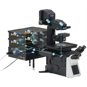
CONFOCAL LASER SCANNING MICROSCOPY
Laser scanning Confocal Microscopy (LSCM) is used in biological research to obtain high-resolution, high-contrast imagery of a sample. Laser microscopes can scan samples point by point, resulting in optical sectioning that can be used to construct precise 3D imagery. Laser scanning microscopes are designed with a large range of imaging modalities to meet some of the most difficult challenges in the life sciences. The FLUOVIEW FV3000 series of confocal laser scanning microscopes meets some of the most difficult challenges in modern science. Featuring the high sensitivity and speed required for live cell imaging as well as deep tissue observation, the FV3000 confocal microscope enables a wide range of imaging modalities, including macro-to-micro imaging, super resolution microscopy, and quantitative data analysis.
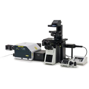
SPINNING DISK CONFOCAL MICROSCOPY (SDCM)
Spinning disk confocal microscopy (SDCM) represents an alternative to LSCM. The Spinning Disk unit helps you acquire high-speed confocal images with a larger field of view than traditional confocal microscopes. Deconvolution algorithms can be applied to improve image resolution, contrast, and dynamic range for strikingly clear 3D images, even at greater observation depths. The Yokogawa CSU-W1 spinning disk unit helps you acquire high-speed confocal images with a larger field of view than traditional confocal microscopes.
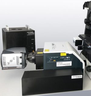

VisiScope Confocal
Real-Time Confocal for wide field of view, improved image quality and high speed live cell imaging